


TIM 3 Recombinant Rabbit Monoclonal Antibody [PSH10-00]
Recombinant Rabbit monoclonal Antibody
Recombinant protein within human TIM 3 aa 1-223.
Human
WB, IF-Cell, FC, IP
Predicted band size: 33 kDa
RPMI 8226 cell lysate, HUT 102 cell lysate, RPMI 8226.
unconjugated
PSH10-00
Liquid
1ug/ul
Store at +4℃ after thawing. Aliquot store at -20℃. Avoid repeated freeze / thaw cycles.
PBS (pH7.4), 0.1% BSA, 40% Glycerol. Preservative: 0.05% Sodium Azide.
IgG
Protein A affinity purified.
WB
1:2,000
IF-Cell
1:100
FC
1:1,000
IP
1-2μg/sample
Hepatitis A virus cellular receptor 2 (HAVCR2), also known as T-cell immunoglobulin and mucin-domain containing-3 (TIM-3), is a protein that in humans is encoded by the HAVCR2 (TIM-3)gene. HAVCR2 was first described in 2002 as a cell surface molecule expressed on IFNγ producing CD4+ Th1 and CD8+ Tc1 cells. Later, the expression was detected in Th17 cells, regulatory T-cells, and innate immune cells (dendritic cells, NK cells, monocytes, macrophages). HAVCR2 receptor is a regulator of the immune response.
1. Kandel S et al. The TIM3/Gal9 signaling pathway: An emerging target for cancer immunotherapy. Cancer Lett. 2021 Jul
2. Palacios LM et al. TIM3 Expression in Anaplastic-Thyroid-Cancer-Infiltrating Macrophages: An Emerging Immunotherapeutic Target. Biology (Basel). 2022 Nov
Membrane, Cell junction, Cell membrane.
CD366 antibody
FLJ14428 antibody
HAVcr-2 antibody
Havcr2 antibody
HAVR2_HUMAN antibody
Hepatitis A virus cellular receptor 2 antibody
Kidney injury molecule 3 antibody
KIM 3 antibody
KIM3 antibody
T cell immunoglobulin and mucin domain containing 3 antibody
展开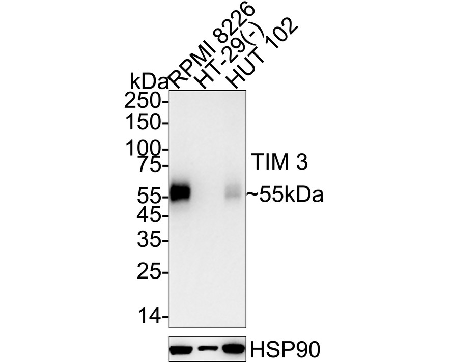
☑ Relative expression (RE)
Western blot analysis of TIM 3 on different lysates with Rabbit anti-TIM 3 antibody (HA723169) at 1/2,000 dilution.
Lane 1: RPMI 8226 cell lysate (20 µg/Lane)
Lane 2: HT-29 cell lysate (negative) (20 µg/Lane)
Lane 3: HUT 102 cell lysate (20 µg/Lane)
Predicted band size: 33 kDa
Observed band size: 55 kDa
Exposure time: 40 seconds; ECL: K1802;
4-20% SDS-PAGE gel.
Proteins were transferred to a PVDF membrane and blocked with 5% NFDM/TBST for 1 hour at room temperature. The primary antibody (HA723169) at 1/2,000 dilution was used in 5% NFDM/TBST at 4℃ overnight. Goat Anti-Rabbit IgG - HRP Secondary Antibody (HA1001) at 1/50,000 dilution was used for 1 hour at room temperature.
☑ Relative expression (RE)
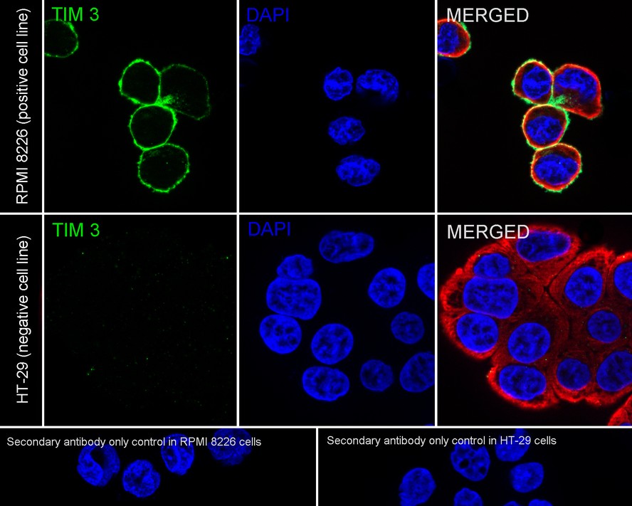
Immunocytochemistry analysis of RPMI 8226 (positive) and HT-29 (negative) labeling TIM 3 with Rabbit anti-TIM 3 antibody (HA723169) at 1/100 dilution.
Cells were fixed in 4% paraformaldehyde for 15 minutes at room temperature, permeabilized with 0.1% Triton X-100 in PBS for 15 minutes at room temperature, then blocked with 1% BSA in 10% negative goat serum for 1 hour at room temperature. Cells were then incubated with Rabbit anti-TIM 3 antibody (HA723169) at 1/100 dilution in 1% BSA in PBST overnight at 4 ℃. Goat Anti-Rabbit IgG H&L (iFluor™ 488, HA1121) was used as the secondary antibody at 1/1,000 dilution. PBS instead of the primary antibody was used as the secondary antibody only control. Nuclear DNA was labelled in blue with DAPI.
Beta tubulin (HA601187, red) was stained at 1/100 dilution overnight at +4℃. Goat Anti-Mouse IgG H&L (iFluor™ 594, HA1126) was used as the secondary antibody at 1/1,000 dilution.
☑ Relative expression (RE)
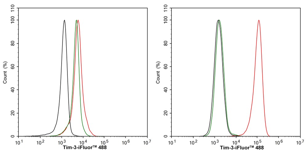
Flow cytometric analysis of HT-29 (left, negative) and RPMI 8226 (right, positive) cells labeling TIM 3.
Cells were fixed and permeabilized. Then stained with the primary antibody (HA723169, 1/1,000) (red) compared with Rabbit IgG Isotype Control (green). After incubation of the primary antibody at +4℃ for an hour, the cells were stained with a iFluor™ 488 conjugate-Goat anti-Rabbit IgG Secondary antibody (HA1121) at 1/1,000 dilution for 30 minutes at +4℃. Unlabelled sample was used as a control (cells without incubation with primary antibody; black).

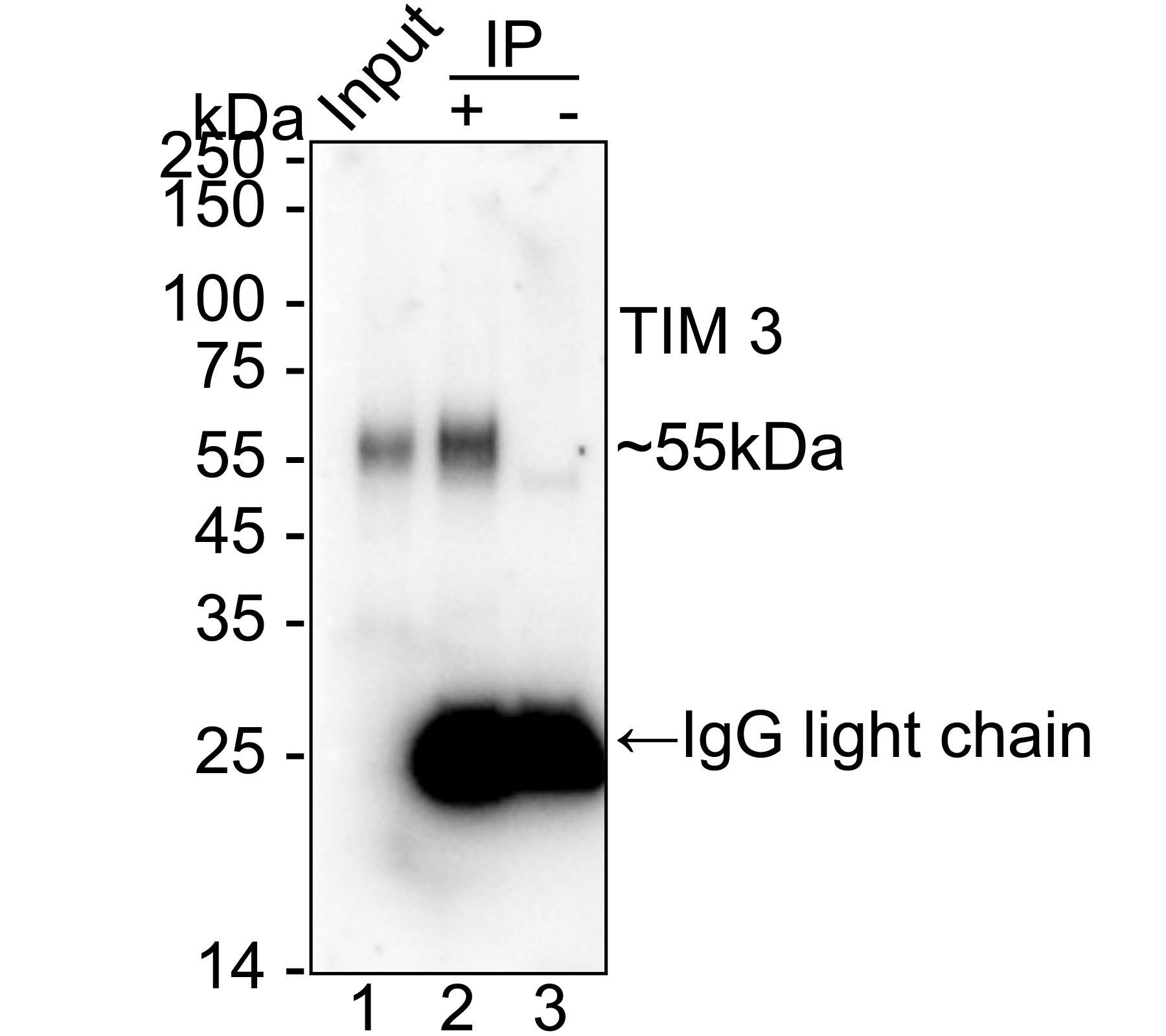
TIM 3 was immunoprecipitated from 0.2 mg RPMI 8226 cell lysate with HA723169 at 2 µg/10 µl beads. Western blot was performed from the immunoprecipitate using HA723169 at 1/2,000 dilution. Mouse Anti-Rabbit IgG kappa light chain secondary antibody (M1208-2) at 1/5,000 dilution was used for 1 hour at room temperature.
Lane 1: RPMI 8226 cell lysate (input)
Lane 2: HA723169 IP in RPMI 8226 cell lysate
Lane 3: Rabbit IgG instead of HA723169 in RPMI 8226 cell lysate
Blocking/Dilution buffer: 5% NFDM/TBST
Exposure time: 59 seconds; ECL: K1802


Copyright © 广州杰特伟生物科技有限公司 All Rights Reserved. 备案号:粤ICP备19077843号