


E-Cadherin Recombinant Rabbit Monoclonal Antibody [SY0287]
Recombinant Rabbit monoclonal Antibody
Synthetic peptide within human E-Cadherin aa 580-630 (Extracellular).
Human (Predicted: Cynomolgus monkey)
WB, IF-Cell, IF-Tissue, IHC-P, FC, IP, mIHC
Predicted band size: 97 kDa
MCF7 cell lysate, HT-29 cell lysate, HCT 116 cell lysate, A431 cell lysate, Caco-2 cell lysate, HT-29, human lung carcinoma tissue, human colon tissue, human breast cancer tissue, human tonsils tissue.
unconjugated
SY0287
Liquid
1ug/ul
Store at +4℃ after thawing. Aliquot store at -20℃ or -80℃. Avoid repeated freeze / thaw cycles.
1*TBS (pH7.4), 0.05% BSA, 40% Glycerol. Preservative: 0.05% Sodium Azide.
IgG
Protein A affinity purified.
WB
1:5,000
IF-Cell
1:2,000
IF-Tissue
1:200
IHC-P
1:200
FC
1:1,000
IP
Use at an assay dependent concentration.
mIHC
1:1,000
| Human | 查看 37 篇文献如下 |
| Mouse | 查看 8 篇文献如下 |
| chicken | 查看 1 篇文献如下 |
Cadherin-1 (not to be confused with the APC/C activator protein CDH1) also known as CAM 120/80 or epithelial cadherin (E-cadherin) or uvomorulin is a protein that in humans is encoded by the CDH1 gene. Mutations are correlated with gastric, breast, colorectal, thyroid, and ovarian cancers. CDH1 has also been designated as CD324 (cluster of differentiation 324). It is a tumor suppressor gene. E-cadherin (epithelial) is the most well-studied member of the cadherin family. It consists of 5 cadherin repeats (EC1 ~ EC5) in the extracellular domain, one transmembrane domain, and an intracellular domain that binds p120-catenin and beta-catenin. The intracellular domain contains a highly-phosphorylated region vital to beta-catenin binding and, therefore, to E-cadherin function. In epithelial cells, E-cadherin-containing cell-to-cell junctions are often adjacent to actin-containing filaments of the cytoskeleton. E-cadherin is first expressed in the 2-cell stage of mammalian development, and becomes phosphorylated by the 8-cell stage, where it causes compaction.
1. Su B et al. Diallyl disulfide suppresses epithelial-mesenchymal transition, invasion and proliferation by downregulation of LIMK1 in gastric cancer. Oncotarget 7:10498-512 (2016).
2. Schmidt TP et al. Identification of E-cadherin signature motifs functioning as cleavage sites for Helicobacter pylori HtrA. Sci Rep 6:23264 (2016).
Non-neural epithelial tissues.
During apoptosis or with calcium influx, cleaved by a membrane-bound metalloproteinase (ADAM10), PS1/gamma-secretase and caspase-3. Processing by the metalloproteinase, induced by calcium influx, causes disruption of cell-cell adhesion and the subsequent release of beta-catenin into the cytoplasm. The residual membrane-tethered cleavage product is rapidly degraded via an intracellular proteolytic pathway. Cleavage by caspase-3 releases the cytoplasmic tail resulting in disintegration of the actin microfilament system. The gamma-secretase-mediated cleavage promotes disassembly of adherens junctions. During development of the cochlear organ of Corti, cleavage by ADAM10 at adherens junctions promotes pillar cell separation (By similarity).; N-glycosylation at Asn-637 is essential for expression, folding and trafficking. Addition of bisecting N-acetylglucosamine by MGAT3 modulates its cell membrane location.; Ubiquitinated by a SCF complex containing SKP2, which requires prior phosphorylation by CK1/CSNK1A1. Ubiquitinated by CBLL1/HAKAI, requires prior phosphorylation at Tyr-754.; O-glycosylated. O-manosylated by TMTC1, TMTC2, TMTC3 or TMTC4. Thr-285 and Thr-509 are O-mannosylated by TMTC2 or TMTC4 but not TMTC1 or TMTC3.
Cell junction, Cell membrane, Endosome, Golgi apparatus.
Arc 1 antibody
CADH1_HUMAN antibody
Cadherin 1 antibody
cadherin 1 type 1 E-cadherin antibody
Cadherin1 antibody
CAM 120/80 antibody
CD 324 antibody
CD324 antibody
CD324 antigen antibody
cdh1 antibody
展开
☑ Relative expression (RE)
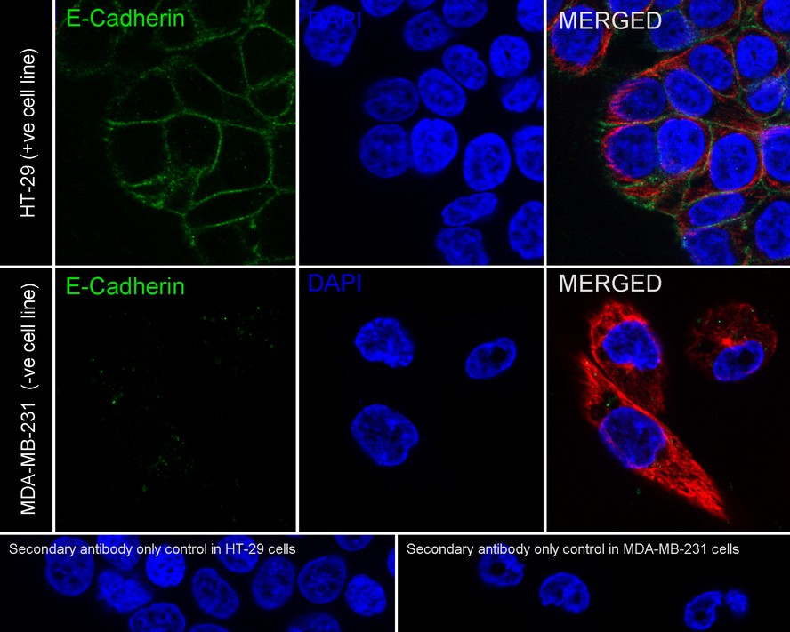
Immunocytochemistry analysis of HT-29 (positive) and MDA-MB-231 (negative) cells labeling E-Cadherin with Rabbit anti-E-Cadherin antibody (ET1607-75) at 1/2,000 dilution.
Cells were fixed in 4% paraformaldehyde for 20 minutes at room temperature, permeabilized with 0.1% Triton X-100 in PBS for 5 minutes at room temperature, then blocked with 1% BSA in 10% negative goat serum for 1 hour at room temperature. Cells were then incubated with Rabbit anti-E-Cadherin antibody (ET1607-75) at 1/2,000 dilution in 1% BSA in PBST overnight at 4 ℃. Goat Anti-Rabbit IgG H&L (iFluor™ 488, HA1121) was used as the secondary antibody at 1/1,000 dilution. PBS instead of the primary antibody was used as the secondary antibody only control. Nuclear DNA was labelled in blue with DAPI.
Beta tubulin (M1305-2, red) was stained at 1/100 dilution overnight at +4℃. Goat Anti-Mouse IgG H&L (iFluor™ 594, HA1126) was used as the secondary antibody at 1/1,000 dilution.

☑ Relative expression (RE)
Western blot analysis of E-Cadherin on different lysates with Rabbit anti-E-Cadherin antibody (ET1607-75) at 1/5,000 dilution and competitor's antibody at 1/1,000 dilution.
Lane 1: MCF7 cell lysate
Lane 2: MDA-MB-231 cell lysate (negative)
Lane 3: HT-29 cell lysate
Lane 4: HCT 116 cell lysate
Lane 5: A431 cell lysate
Lane 6: Caco-2 cell lysate
Lysates/proteins at 20 µg/Lane.
Predicted band size: 97 kDa
Observed band size: 80~120 kDa
Exposure time: 43 seconds; ECL: K1801;
4-20% SDS-PAGE gel.
Proteins were transferred to a PVDF membrane and blocked with 5% NFDM/TBST for 1 hour at room temperature. The primary antibody (ET1607-75) at 1/5,000 dilution and competitor's antibody at 1/1,000 dilution were used in 5% NFDM/TBST at 4℃ overnight. Goat Anti-Rabbit IgG - HRP Secondary Antibody (HA1001) at 1/50,000 dilution was used for 1 hour at room temperature.

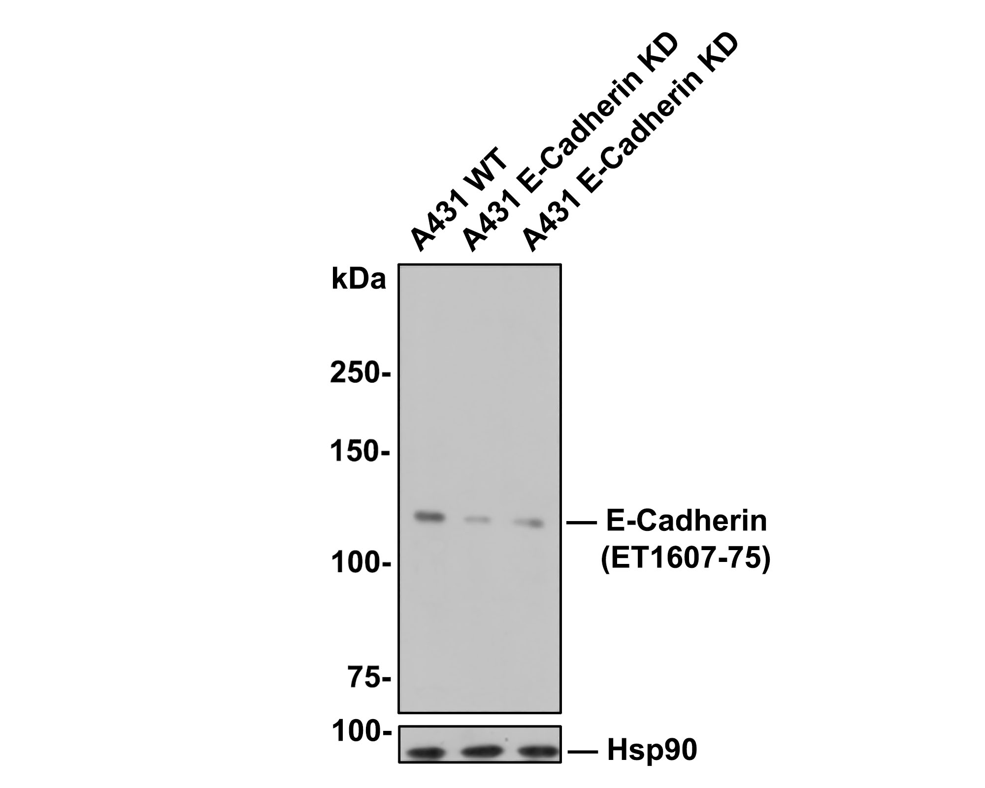
☑ Knockdown (KD)
Western blot analysis of E-Cadherin with anti-E-Cadherin antibody (ET1607-75) at 1/5,000 dilution.
Lane 1: Wild-type A431 whole cell lysate (10 µg).
Lane 2/3: E-Cadherin knockdown A431 whole cell lysate (10 µg).
Proteins were transferred to a PVDF membrane and blocked with 5% NFDM in TBST for 1 hour at room temperature. The primary antibody (ET1607-75, 1/5,000) was used in 5% BSA at room temperature for 2 hours. Goat Anti-Rabbit IgG-HRP Secondary Antibody (HA1001) at 1/100,000 dilution was used for 1 hour at room temperature.
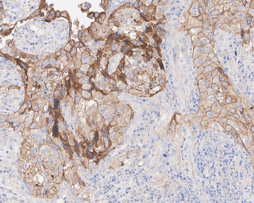
Immunohistochemical analysis of paraffin-embedded human lung carcinoma tissue with Rabbit anti-E-Cadherin antibody (ET1607-75) at 1/200 dilution.
The section was pre-treated using heat mediated antigen retrieval with Tris-EDTA buffer (pH 9.0) for 20 minutes. The tissues were blocked in 1% BSA for 20 minutes at room temperature, washed with ddH2O and PBS, and then probed with the primary antibody (ET1607-75) at 1/200 dilution for 1 hour at room temperature. The detection was performed using an HRP conjugated compact polymer system. DAB was used as the chromogen. Tissues were counterstained with hematoxylin and mounted with DPX.

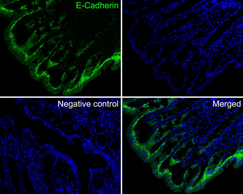
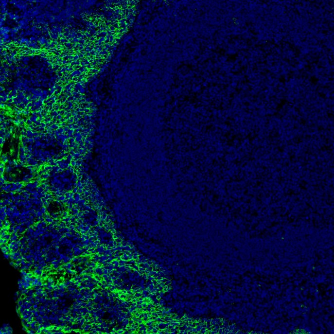
Application: IF-tissue
Species: Human
Site: Colon
Sample: Paraffin-embedded section
Antibody concentration: 1/200
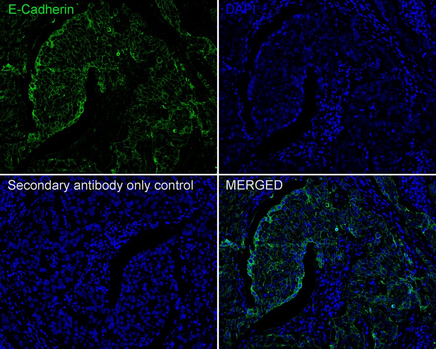
Application: IF-tissue
Species: Human
Site: Breast cancer
Sample: Paraffin-embedded section
Antibody concentration: 1/200
mIHC analysis of human tonsils tissue (Formalin/PFA-fixed paraffin-embedded sections) with Rabbit anti-E-Cadherin antibody (ET1607-75) at 1/1,000 dilution. The immunostaining was performed with the IRISKit® HyperView mTSA Kit (MH900206). Heat mediated antigen retrieval with Tris-EDTA buffer (pH 9.0) for 30 mins at 95℃. DAPI (blue) was used as a nuclear counter stain. Image acquisition was performed with Olympus VS200 Slide Scanner.
☑ Relative expression (RE)
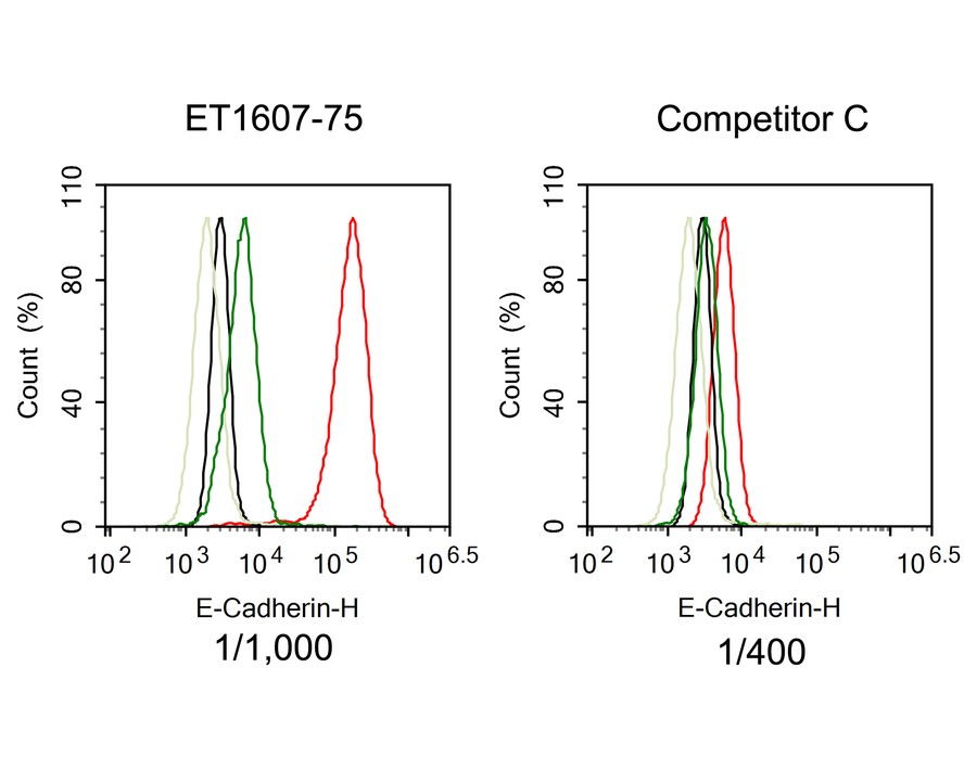
Flow cytometric analysis of HT-29 (positive, red) and MDA-MB-231 (negative, green) cells labeling E-Cadherin.
Cells were fixed and permeabilized. Then stained with the primary antibody (ET1607-75, red) at 1/1,000 dilution and competitor's antibody (red) at 1/400 dilution, compared with Rabbit IgG Isotype Control (HT-29 black, MDA-MB-231 light green). After incubation of the primary antibody at +4℃ for an hour, the cells were stained with a iFluor™ 488 conjugate-Goat anti-Rabbit IgG Secondary antibody (HA1121) at 1/1,000 dilution for 30 minutes at +4℃.
Decoupling of Density-Dependent Migration/Proliferation Dichotomy on Surface Potential Gradient
Author: Zejun Chen, Lingqing Dong
PMID: 40036071
期刊: ACS Applied Materials & Interfaces
应用: IF,WB
反应种属: Human
发表时间: 2025 Mar


Copyright © 广州杰特伟生物科技有限公司 All Rights Reserved. 备案号:粤ICP备19077843号